Intraoral Scan – Digital Impressions with Planmeca Primescan in Bangalore
In our digital dentistry workflow, the old-fashioned dental impression is no longer the part you dread most. Instead of a metal tray loaded with cold, gooey material that sits in your mouth for minutes, which can trigger a gag reflex and may need to be repeated if you move, we now capture your teeth digitally. Traditional impressions then have to be poured in stone, trimmed, and handled in the lab, with tiny changes from material shrinkage, expansion, and distortion adding up to affect how precisely your final crown fits.
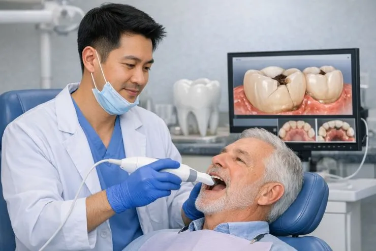
The Planmeca Primescan intraoral scanner replaces this entire process. A small, lightweight wand is passed gently over your teeth while a structured light system captures a continuous stream of 3D surface data in real time, building a precise digital model of your teeth and gums on the chairside screen: no impression material, no trays, no gagging, and no waiting. The scan is checked immediately, any area needing more detail is rescanned on the spot, and the complete digital model is sent to the lab or milling unit within minutes.
At Dental Solutions Clinic in Indiranagar, Bangalore, Planmeca Primescan is the standard impression method for restorative, cosmetic, implant prosthetic, and orthodontic work. It is used primarily by Dr. Ramya Balasubramanya – MDS Prosthodontics, BDS Gold Medallist, Certified DSD Practitioner, Certified Invisalign Provider – and is fully integrated into our DSD smile design workflow, CAD/CAM fabrication, and the Invisalign ClinCheck treatment planning platform.

What Is an Intraoral Scanner?
An intraoral scanner is a handheld device that records the 3D shape of your teeth, gums, and bite using structured light, laser, or confocal imaging. It projects a light pattern onto the tooth surface, a camera captures how that pattern bends over each contour, and software converts this into precise 3D coordinates to build a continuous digital model in real time. The result is a 3D mesh of your teeth – a digital impression in STL or PLY format – typically accurate to within a few tens of microns across a full arch.
How We Use Intraoral Scans
- Crown and veneer impressions
Replaces conventional impressions for all CAD/CAM restorations, capturing the prepared tooth, margins, and adjacent teeth in a single scan and sending the model straight to the design software. - Bridge and implant impressions
Records all abutment preparations and implant scan bodies, along with their exact spatial relationships, so that multi-unit bridges and implant restorations can be planned and fabricated with high accuracy. - Digital Smile Design (DSD)
The intraoral scan supplies the 3D tooth data that is merged with DSD facial photographs; proposed veneers are designed directly on the digital model and then matched to the facial overlay to create a complete digital brief for the lab. - Invisalign and orthodontic records
The scan fully replaces alginate impressions for Invisalign; data is uploaded directly to the Invisalign cloud, where the ClinCheck simulation is generated without the need to ship models. - Full-mouth rehabilitation records
Full-arch scans document existing tooth positions, wear, and bite, supporting digital occlusal analysis, articulated model evaluation, and fabrication of provisional and final restorations across both arches. - Night guard and splint fabrication
A full-arch scan replaces traditional impressions for night guards, splints, and study models, eliminating material distortion and storage of plaster casts. - Study models and monitoring
Periodic scans during orthodontic or restorative review enable digital comparison of models over time, accurately tracking tooth movement and arch development without the need for physical models.
The Scanning Process – What to Expect
An intraoral scan at DSC usually takes 2–8 minutes, depending on how much we are scanning: about 2 minutes for a single tooth and 5–8 minutes for both arches with bite.
Preparation
The scanner tip, covered with a single-use sleeve, is gently warmed to reduce fogging, and the monitor is positioned so you can watch the 3D model build as we scan.
Scanning sequence
We start on the biting surfaces of the back teeth and move forward in a defined pattern, then capture the cheek (buccal) and tongue (lingual) sides, scanning each surface from multiple angles to avoid shadows or gaps.
Real-time review
The 3D model builds live on-screen; any areas with insufficient data – such as under gum margins or between tight contacts – are highlighted and rescanned immediately, without starting over.
Bite registration
For cases involving your bite, we record how the upper and lower teeth meet by scanning while you close together naturally, replacing the old bite registration wafer.
Transmission
Once approved, the model is exported as an STL file and sent straight to CAD/CAM design software, the lab portal, or uploaded to the Invisalign ClinCheck platform – typically within 2 minutes of finishing the scan.
Digital vs Conventional Impressions
| Aspect | Conventional impression | Digital impression at DSC |
|---|---|---|
| Patient comfort | Trays, gagging risk, 3–5 minutes of material | Small wand, no material, no gagging |
| Accuracy – single tooth | Good, but material- and technique-dependent | Excellent, consistently tight marginal fit |
| Accuracy – full arch | Falls with arch length due to material distortion | Maintained across the arch, full-arch accuracy in tens of microns |
| Retake rate | Higher, tears or poor margins are common | Lower, gaps seen and rescanned in real time |
| Lab transit time | 1–3 days for model transport | Minutes, files sent instantly |
| Storage | Bulky stone models, risk of damage or loss | Compact digital files, no physical storage needed |
| CAD/CAM integration | Requires model scanning, adds another error step | Direct digital input to CAD/CAM |
| Invisalign compatibility | Physical models must be shipped or scanned | Direct cloud upload to ClinCheck, no shipping or delay |
Digital impressions with intraoral scanners consistently show higher patient comfort and at least comparable, often superior, accuracy to conventional techniques, especially in real clinical conditions.
Frequently Asked Questions
Is an intraoral scan as accurate as a conventional impression?
Yes. For single crowns, veneers, and short bridges, high-quality intraoral scanners are at least as accurate as conventional impressions and often more so, with full-arch scans also achieving clinically acceptable accuracy when used correctly. Proper technique, retraction, and complete margin capture are essential for the best results.
I have a strong gag reflex. Will an intraoral scan be better for me?
Almost always. Gagging with conventional impressions is mainly caused by impression material and bulky trays at the back of the mouth, whereas the scanner uses a slim wand and no setting material, so most gag-prone patients tolerate it much more easily.
Can children have an intraoral scan?
Yes. Children usually accept the small scan wand far better than full trays, making digital scans ideal for orthodontic records, study models, and space maintainers while avoiding the discomfort and waiting time of traditional alginate impressions.
Do I need an intraoral scan for Invisalign?
Yes. Invisalign’s ClinCheck planning is based on a digital scan of your teeth, captured at DSC with the intraoral scanner and uploaded to create a 3D model and a step-by-step tooth-movement simulation used to fabricate your aligners. No physical impressions or model shipping are needed.
Does an intraoral scan replace the need for X rays?
No. Scans record the 3D surface shape of teeth and gums, while X-rays show internal tooth and bone structures, decay, and deeper anatomy; both are needed for complete diagnosis and planning, and they complement each other in a modern digital dentistry workflow.