RVG Digital X-Ray — Low-Radiation Digital Dental Radiography in Bangalore
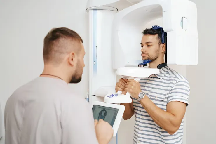
As part of our fully integrated digital dentistry workflow, RVG (Radiovisiography) replaces old film-based X-rays with fast, low-radiation, sensor-driven imaging at the chairside. Instead of waiting minutes for films to develop, a high-resolution digital X-ray appears on the monitor in a few seconds, with a radiation dose up to 80% lower than conventional film. This image drops straight into your digital record, can be enhanced and measured on screen, and links seamlessly with our CBCT scans, intraoral scans, and CAD/CAM planning for more precise diagnosis and treatment.
The benefit is more than convenience. Digital radiographs can be zoomed, contrast‑adjusted, and measured precisely on screen without damaging the original data, helping clinicians spot small changes in bone, early decay, or fine root details that might be missed on fixed‑contrast film. At Dental Solutions Clinic in Indiranagar, Bangalore, RVG digital X-rays are the standard for routine check-ups, restorative and periodontal planning, root canal assessment, and implant follow-up, forming a core part of our diagnostic and monitoring process.
What Is RVG (Radiovisiography)?
RVG is a digital intraoral X‑ray system that replaces conventional film with a small electronic sensor placed inside the mouth. The sensor (CCD or CMOS) converts X‑ray photons directly into a digital signal, which appears as a high‑resolution greyscale image on the computer within 2–3 seconds. Because these sensors are more sensitive than film, diagnostic‑quality images can be captured with significantly lower radiation exposure.
Developed in the late 1980s and now widely used in modern practice, RVG has become the standard intraoral radiography method in many specialist clinics due to its speed, dose reduction, and image-enhancement capabilities.
When We Use RVG Digital X-Rays
Digital RVG imaging is used across disciplines at Dental Solutions Clinic, with different views chosen depending on the clinical question:
- Periapical X‑rays – show the whole tooth and surrounding bone, used for root canal diagnosis, apical infections, fractures, and post‑treatment checks.
- Bitewing X‑rays – show the crowns of upper and lower back teeth together, ideal for detecting cavities between teeth and monitoring bone levels under old fillings and crowns.
- Full‑mouth periapical series – a set of images covering all teeth for comprehensive periodontal, restorative, or full‑mouth rehabilitation planning.
- Occlusal X‑rays – wider views of the palate or floor of the mouth for impacted teeth or certain jaw conditions.
- Post‑operative periapicals – taken immediately after root canals, extractions, or implant placement to confirm lengths, positions, and initial healing.
What RVG Helps Us Diagnose

Early cavities between teeth
Digital bitewings help detect interproximal decay at a stage when small, minimally invasive fillings or remineralisation strategies are still possible.

Periodontal bone loss
Periapical series allows precise measurement of bone height at each tooth, supporting accurate staging and monitoring of gum disease treatment.

Root canal length and anatomy
Sequential RVG images during root canal treatment confirm working length and filling quality, improving predictability and reducing chair time.

Implant monitoring
Periapical RVGs are used to check implant positioning, integration, and crestal bone levels over time with measurable accuracy.
Radiation Safety
Typical effective doses for intraoral dental X‑rays are very low — around 1–8 microsieverts (µSv) per periapical or bitewing image, which is comparable to just a few hours of natural background radiation. Digital RVG systems can reduce this exposure by roughly 50–80% compared with film, as sensors require less radiation to produce a clear image.
At Dental Solutions Clinic, every X‑ray is taken only when clinically justified, following the ALARA principle (As Low As Reasonably Achievable). Protective lead aprons and thyroid collars are available for all patients who request them.

Frequently Asked Questions
Is a dental X ray safe?
Yes. Digital RVG X rays use a very low dose — roughly equivalent to less than an hour of natural background radiation for a single small image, and around 80% less than old film X rays. The benefit of early, accurate diagnosis far outweighs this minimal exposure.
Do I need an X ray at every visit?
No. X rays are taken only when there is a clear clinical reason, such as checking for decay between teeth, assessing pain, or planning treatment. High risk patients may need bitewings more often; low risk patients may only need them every few years.
Why does the image appear so quickly?
The RVG sensor converts X ray energy directly into a digital image, so there is no film or chemical developing step. The picture appears on the screen within a few seconds.
Can you adjust the image after taking it?
Yes. Digital X rays can be zoomed, lightened or darkened, and contrast enhanced without harming the original data, helping us see fine details such as early decay or subtle bone changes more clearly.
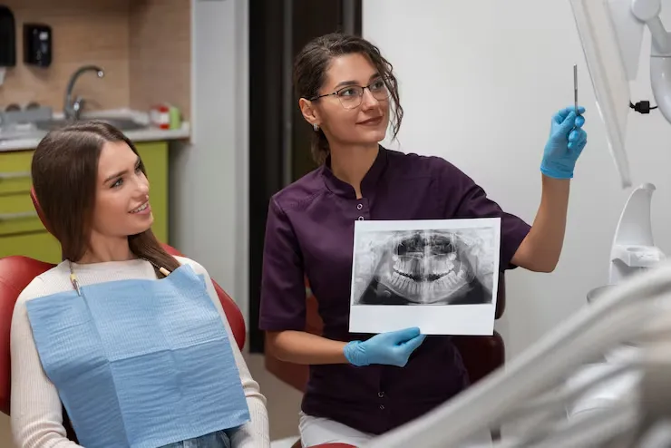
What is the difference between an RVG and an OPG?
RVG is an intraoral sensor used for detailed views of one or a few teeth (periapical or bitewing images). An OPG is a panoramic scan taken from outside the mouth that shows all teeth, jaws, and joints in one overview, but with less fine detail per tooth. Both are digital; each is chosen for different diagnostic needs.
Are X rays done differently for children?
We use the same RVG system with smaller sensors for children and adjust exposure for their thinner jaws, so the dose is as low as reasonably achievable. X rays are taken only when they are likely to change or guide treatment.